China World Technology Medical Equipment Service Group
Win-win together!
 |
| Certification: | CE |
| Model Number: | C6-2 |
| Minimum Order Quantity: | 1pcs |
|---|---|
| Packaging Details: | Carton |
| Delivery Time: | 3-5 working days |
| Payment Terms: | T/T(wire transfer), Western Union, Paypal |
| Brand Name: | Philip | Product Name Q: | C6-2 Curved Array Philip Affiniti 50G Ultrasound Probe Repair 2.0 MHz. -6.0 MHz |
|---|---|---|---|
| Item No.: | C6-2 | Technology: | Broadband |
| Frequency Range: | 6-2 MHz | Number Of Elements: | 128 |
| Aperture: | 63.7 Mm | Array Type: | Curved |
| Applications: | General Purpose Abdominal (adult And Pediatric, Including Vascular), Bowel, Obstetrical, Gynecological, Prostate And Interventional Applications | Modes: | 2D, Steerable Pulsed, High-PRF, And Color Doppler, Color Power Angio (CPA), Directional CPA, SonoCT, XRES And Multivariate Harmonic Imaging |
| Field Of View: | 95° | ||
| High Light: | Affiniti 50G Ultrasound Transducer Probe,6.0 MHz Ultrasound Transducer Probe,Curved Array Ultrasound Transducer |
||
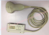
C6-2 Curved Array Philipilip Affiniti 50G Ultrasound Transducer Probe Repair 2.0 MHz. -6.0 MHz
The frequency range of the Philipilip C6-2 curved array transducer is 2.0 MHz. -6.0 MHz. And supports the following applications: abdomen, obstetrics, gynecology, anesthesia, nerves, pelvis.
Description
| 63.7 mm |
| Applications | General purpose abdominal (adult and pediatric, including vascular), |
| Technology | Broadband |
| Number of elements | 128 |
| Image Fusion Navigation capable | No |
| Array Type | Curved |
| Biopsy capable | Yes |
| Frequency range | 6 - 2 MHz |
| Modes | 2D, steerable pulsed, High-PRF, and color Doppler, |
| Field of view | 95° |
| Volume of field of view | / |
| Philipilipysical dimensions | / |
Common faults and causes of Philipilip ultrasonic probe waves:
Ultrasound probes often fail due to frequent use and direct contact with the subject, which affects the normal use of the hospital. Many hospitals will give priority to probe failures
01 Acoustic lens and matching layer failure
Due to long-term direct contact with human skin, mucous membranes or the outside world, the acoustic lens is susceptible to impact, scratches, abrasion, corrosion, cracking, degumming, blistering, etc., which will affect the image diagnosis effect; if the acoustic lens and matching layer are completely peeled off, the chip If exposed, there may be a risk of electric shock to the patient. At the same time, after the exposed chip comes into contact with the coupling agent or disinfectant, it is easy to corrode the chip, resulting in damage to the crystal that cannot be repaired; therefore, it needs to be repaired or replaced with a new probe immediately.
Common causes of failure:
The acoustic lens is in direct contact with the human body for a long time. If the human body has more body hair, it will accelerate the wear;
The operator has not developed the habit of wiping clean the surface coupling agent or disinfectant after using the probe;
The plastic and rubber materials of the acoustic lens and the matching layer are affected by temperature. For example, a large temperature difference will cause the thermal expansion and contraction to cause degumming, while accelerating aging and making it more susceptible to deformation and cracking.
02 Crystal failure
Crystal failure, often manifested in the image as attenuation, dark shadows, bright white bands, missing images, etc., in severe cases, the overall image "blindness"
Common causes of failure:
03 Circuit board failure
Circuit board failure usually manifests as no image or image defect during inspection.
Common causes of failure:
![]()
04 Sheath repair
The sheath is located at the junction of the cable and the probe shell. The sheath can protect the cable to prevent the junction of the cable and the shell from breaking and breaking under the condition of frequent bending. If the sheath is cracked or damaged, not only will the EMC performance of the probe be degraded, which will cause the image to produce noise, but also cause the image to be dark or missing.
Common causes of failure:
Because the probe needs to examine the patient from different angles, the cable sheath at the root of the probe aggravates fatigue fracture, and the cable loses its protective layer, resulting in damage to the cable.
05 Cable fault
Generally, it shows that the protective layer and shielding layer of the cable are broken or even broken. In severe cases, it is reflected in the image, where part of the image is missing or noise interference, system crashes, and probes do not recognize faults.
Common causes of failure:
During long-term use, the cable, especially the cable close to the sound head, is always in a state of bending and torsion;
The operator's unintentional pulling operation: When inspecting the far part, instead of shortening the distance, the cable is pulled directly; or when using a cart, the cover of the probe cable is pressed by the wheels of the cart.
06 Shell repair
Due to the destruction of the shielding quality of the probe shell, the image will be disturbed and unclear. In severe cases, an induced current will appear from the front end, which will endanger the patient's body.
Common causes of failure:
Long-term use of the probe will cause cracking and aging of the shell, or deformation due to man-made factors such as falling or touching.
Tips on probe maintenance
1. Handle with care during use to avoid collision; after using the probe, the connecting wire of the probe should not be folded or tangled.
2. When plugging or unplugging the probe, make sure that the probe is not in working condition or the power has been turned off.
3. After the probe is used, it should be straightened and hung and the coupling agent should be wiped clean in time. To wipe the probe, clean it with a soft cloth dipped in cleaning fluid (such as neutral soapy water), then use a soft cloth dipped in water to remove the cleaning fluid, and then wipe it dry with a clean soft dry cloth. Do not use alcohol or alcohol-containing wipes for the probe, otherwise it may cause the probe cable or bushing to harden.
4. When disinfecting the ultrasonic probe, use a special liquid chemical disinfectant for disinfection, such as glutaraldehyde solution and disinfectant solution. After disinfection, clean the disinfectant on the probe with sterile water, and then wipe the probe dry with a disinfected soft dry cloth. Remember that the immersion part of the probe should not exceed the azimuth mark on the side of the probe housing, and the immersion time of the probe should not exceed 1 hour. High temperature and high pressure can not be used for disinfection, it will cause damage to the internal structure of the probe.
Contact Person: Kiara
Tel: 8619854815217